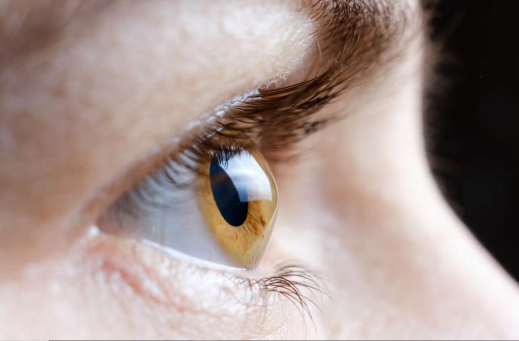
Fuchs Endothelial Dystrophy (FED) is a progressive eye disorder that affects the cornea, the transparent, dome-shaped tissue at the front of the eye. It primarily affects the endothelial cells, a thin layer of cells which maintain corneal clarity and regulate the balance of fluid. We are born with a certain number of endothelial cells and lose them at a slow rate throughout our lives. No new endothelial cells are generated, and when too many of these cells are lost or dysfunctional, the cornea begins to swell and turns cloudy. As a result, light cannot pass through as clearly, leading to blurred or distorted vision. Although the condition is not widely known, its effects on eye health can be significant if not managed appropriately.
Symptoms of Fuchs’ Endothelial Dystrophy
The symptoms of FED can vary from person to person and often develop gradually. The condition usually affects both eyes and manifests later in life, often over the age of 50. Common signs and symptoms include:
- Blurry or Hazy Vision: The corneal swelling can cause images to appear blurry, especially in the morning after waking up, and vision can slowly improve during the day. This is due to fluid accumulation in the cornea overnight.
- Halos Around Lights: The swelling of the cornea can distort the way light enters the eye, leading to the appearance of halos, particularly in low-light conditions.
- Increased Sensitivity to Light: As the condition progresses, individuals with FED may find bright lights uncomfortable and experience glare, which can interfere with everyday activities like driving at night.
- Eye Pain or Discomfort: Some people with Fuchs’ Endothelial Dystrophy may experience pain or gritty sensation.
- Declining Vision Over Time: Without intervention, the swelling can worsen, leading to a more significant loss of vision, and eventually causing legal blindness.
Causes and Risk Factors
While FED has a hereditary component, it often occurs sporadically without any family history. The severity and onset of symptoms can vary greatly between individuals.
Some common risk factors associated with FED include:
- Age: People with FED typically do not develop symptoms until over the age of 50, though it can appear earlier in some cases.
- Family History: If someone in your family has been diagnosed with FED, your chances of having it increase.
- Gender: Women are more likely to develop FED than men, and the condition may also progress more rapidly in females.
- History of Intraocular Surgery: Previous eye surgeries, such as cataract surgery, may accelerate endothelial cell loss and worsen FED symptoms.
Diagnosis of Fuchs’ Endothelial Dystrophy
Diagnosing FED requires a comprehensive eye exam by an ophthalmologist. A few key tests and evaluations include:
- Slit Lamp Examination: This test allows the doctor to closely examine the structure of the eye, including the cornea. They may notice signs of corneal swelling or changes in the endothelial cells.
- Specular Microscopy: This non-invasive test uses a special microscope to take detailed images of the corneal endothelial cells. It helps assess how many healthy cells remain and whether they are functioning properly.
- Corneal Thickness Measurement (Pachymetry): This test measures the thickness of the cornea, which can become thicker as fluid accumulates in FED.
- Visual Acuity Test: This standard eye test measures how well you can see at various distances and can help track the progression of visual impairment.
Treatment Options for Fuchs’ Endothelial Dystrophy
While there is no cure for FED, several treatment options can help manage the symptoms and slow the progression of the disease. The choice of treatment largely depends on the severity of the condition.
Non-surgical treatment options such as hypertonic saline eye drops are used for symptomatic FED to reduce corneal swelling and as a temporising measure ahead of definitive surgical treatment. The drops work by drawing excess fluid out of the cornea, helping to relieve discomfort and blurriness caused by oedema.
Treatment of advanced FED involves surgical intervention to replace the dysfunctional endothelial cell layer with transplanted healthy donor endothelial tissue. Depending on the stage of the FED and the health of the surrounding ocular tissue, your surgeon may recommend the following types of corneal graft surgery:
- Descemet’s Stripping Automated Endothelial Keratoplasty (DSAEK): In cases that are more advanced and where the corneal clarity is significantly impaired, a corneal transplant like DSAEK may be recommended. During this procedure, the dysfunctional endothelial cell layer is removed, and a partial thickness donor cornea tissue is transplanted to restore function.
- Descemet Membrane Endothelial Keratoplasty (DMEK): This is an advanced and precise form of corneal transplant that involves removing the dysfunctional endothelial cell layer and replacing it with a similar thin layer of tissue containing healthy donor endothelial cells. DMEK is suitable for less advanced cases of FED and offers advantages such as faster recovery times and better outcomes.
Living with Fuchs’ Endothelial Dystrophy
While FED can be a challenging corneal disease, many individuals can manage it effectively with appropriate treatment. Regular visits to an ophthalmologist are essential for monitoring the progression of the disease and adjusting treatment as necessary.
In cases where more advanced treatments are needed, corneal transplantation has a high success rate, with many patients achieving significant improvement in vision.Early detection is key to successful management, so regular eye exams are essential for those at risk, particularly individuals with a family history or other risk factors.
Original Source https://nexuseyecare.com.au/eyes/what-is-fuchs-endothelial-dystrophy/

I am really inspired along with your writing skills as well as with the layout in your weblog. Either way keep up the nice high quality writing, it’s uncommon to peer a great blog like this one today.
I really treasure your piece of work, Great post.